Benefits of Using Ultrasound to Diagnose Musculoskeletal Issues
While doctors can learn a lot from a talking with a patient and conducting a physical exam, it often helps to get a look inside the body to see what’s causing discomfort, immobility or other symptoms. X-rays and MRI scans can be useful tools, but they have their limitations. Another type of imaging – ultrasound – offers some distinct advantages.
When we think of ultrasound, we often think of prenatal visits in which an expectant mother gets a first glimpse of her baby, but the technology has multiple applications across many types of medicine, including orthopedics. Ultrasound imaging offers a number of benefits in diagnosing musculoskeletal issues, including arthritis, sports injuries, rotator cuff injuries, tendonitis, bursitis and more. And its uses aren’t just limited to diagnostics. Ultrasound sometimes is used in certain treatments, such as injection therapies and biopsies, to help target treatment to a precise location.
According to Palmetto Bone & Joint’s Physical Medicine and Rehabilitation specialist, Dr. Alaric Van Dam, there are a number of reasons ultrasound is uniquely suited for use in orthopedics:
1. Ultrasounds are remarkably safe.
Ultrasound, also known as sonography, uses sound waves to develop images of body tissues. No dyes or anesthetics are needed. And unlike X-rays or CT scans, there’s not even a low dose of radiation exposure. In fact, there are no known side effects of the procedure.
2. Ultrasound captures images of soft tissues.
X-rays are excellent tools for viewing bones, and orthopedists can make inferences about what’s going on with the soft tissues around those bones based on what they see in an X-ray image. But to actually see those tissues, you need an imaging tool that’s more sensitive. Ultrasound imaging captures muscles, tendons, ligaments, nerves and cartilage.
3. Ultrasound works in different positions.
Standard X-ray or MRI machines require a patient to lie down, stand or sit up in a specific position. Lying down for an imaging procedure only captures what your body is doing when you’re lying down.
4. Ultrasound can see what happens when you move.
Some imaging techniques only work when the patient holds still, so only captures static images. With ultrasound, the patient can move during the procedure, so the doctor or technician can see how tissues act or react to certain movement, such as bending, twisting or reaching, for instance. This can be crucial for diagnosing symptoms that affect mobility or that only occur when you’re moving.
5. Ultrasound offers more accurate diagnosis.
Ultrasound produces images in real time and show doctors a much more accurate picture of what occurs in the body during movement. This translates to more accurate diagnosis – and more accurate treatment for patients.
What to Expect During an Ultrasound Procedure
An ultrasound procedure is relatively simple. A doctor or technician will apply a small amount of gel to your skin. He or she then will use an instrument called a transducer, which glides across the skin. The transducer emits sound waves and sends images to a computer based on what those sound waves detect. The doctor may ask you to move your limbs or joints in certain ways during the procedure, so he or she can see what’s happening during those movements.
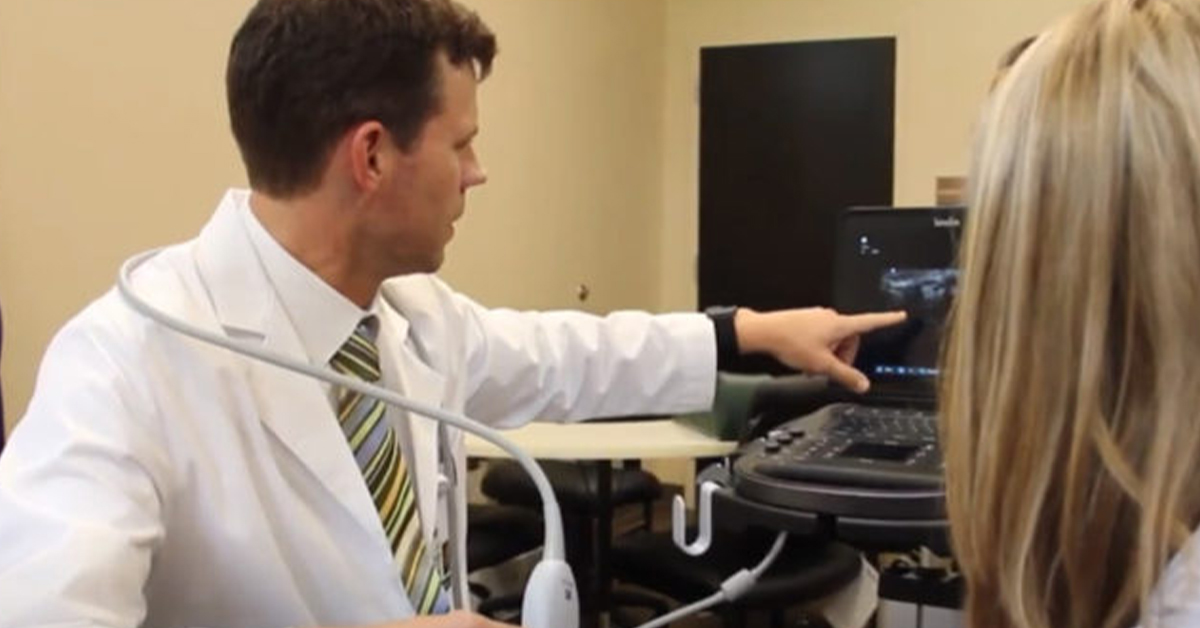
At Palmetto Bone & Joint, ultrasound is one of many tools we use every day to diagnose and treat patients. Learn more as Dr. Van Dam talks about the advantages of ultrasound technology:
Palmetto Bone & Joint is Here for You
Have a bone or joint injury or problem you’d like us to evaluate? Please don’t hesitate to contact our office today!


